What is the Late Anatomy Scan?
The late anatomy scan is a comprehensive ultrasound examination, typically performed between weeks 21–24 of pregnancy. The purpose of the examination is to systematically evaluate the fetal structure and development of body systems at a more advanced stage of pregnancy.
The examination includes assessment of the brain, heart, spine, face, limbs, kidneys, digestive system and additional organs. The scan enables identification of structural findings that may appear or become clearer only later in pregnancy.
Additionally, it's possible to assess fetal growth, amniotic fluid volume, placental location and sometimes perform blood flow evaluation according to clinical need.
Dr. Charak performs the scan with a professional, thorough and sensitive approach, using advanced ultrasound equipment that enables high-quality and clear imaging. During the examination, emphasis is placed on clear, patient and personalized explanation, to provide parents with a sense of confidence and complete understanding.
Fetal Heart — Central Focus of the Scan
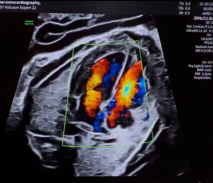
Fetal heart assessment is one of the most complex and important challenges in the late systems review. Dr. Charak performs a comprehensive echocardiographic examination of the heart structure using advanced imaging techniques.
Four chambers of the heart: examination of size, position and symmetry
Heart valves: mitral, tricuspid, aortic and pulmonary
Origin of the great vessels: aorta and pulmonary artery
Aortic arch and Doppler blood flows
Partitions between ventricles and between atria
Frequently Asked Questions
Schedule Your Late Review
Contact Life Inside clinic in Raanana to schedule an appointment with Dr. Ron Charak. We are here to answer any question and provide you with peace of mind throughout pregnancy.